
LOCOMOTION IN PROTOZOA
INTRODUCTION
- Moving ones body is an essential need of any organism. Some organism are sessile while other moves at very high speed. Some organisms moves very slowly such as amoeba or euglena where as other can move fastest example leopard. There is a vast diversity of organism on the basis of their locomotion. In class protozoa classification is done on the basis of its locomotory organ only. Imagine if there is such a much difference in one phylum how much difference must be present in whole animal kingdom.
Some of the time locomotion is misunderstood with the term movement, locomotion is define as movement of whole body of an organism from one place to another hence it can said that locomotion is a kind of movement in which whole body moves where as in movement it is not necessary that whole body move only a single part can also move example peristaltic movement of intestine.
AMOEBOID MOVEMENT
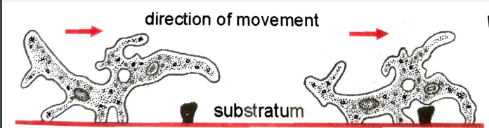
Amoebiod movement is shown by Amboeba by the formation of pseudopodia which is finger like projection. These are broad with a round tip and are called lobopodia. They are formed by the flow of cytoplasm in forward direction.
Theories of amoeboid movement
How the pseudopodia is formed is still not very clear various theories are put forward to explain same but there no sufficient evidence to proof any hypothesis correct.
[a] Contractile hydraulic theory
- It was proposed by Schultze in 1875. It states that ectoplasm also known as plasmagel undergoes contraction at the posterior end and causes protoplasmic current to flow forward, pushing the more fluid like endoplasm also known as plasmasol forward. This results in the formation of pseudopodium and pushing the body forward.
[b] Surface tension theory
- It was proposed by Berthold in 1886. It states that there is difference in the surface tension between the physical characteristics of the body and substratum which results in amoeboid movement. In this theory amoeboid movement is compared with movement of fluid globule mercury droplet. A pseudopodium is formed by the outflow of protoplasm also known as fountain streaming from external and internal factors.
- This theory assume the liquid form of the body surface but in majority of amoeboid form it is rigid and gelatinous.
[c]Rolling movement theory
- This theory was given by Jennings in 1904. He did his studies on Amoeba verrucosa. He compare amoeboid movement with rolling movement of fluid filled sac on substratum.
- He observed that a carbon particle on amoebas upper surface first passes forward and then turn downward on anterior tip, remains on lower surface for a time as body rolls forward and then passes upwards at the posterior end to repeat this cycle.
- His finding may be correct for Amoeba verrucosa which is devoid of pseudopodia but it cannot applied to A. Proteus which is devoid of pseudopodia.
[d] Walking movement theory
- This theory was proposed by Dellinger in year 1906. He did his studies on A. Proteus and conclude that there is presence of contractile substances which are mainly responsible for amoeboid movement.
- According to this theory the extended pseudopodia attached to substratum and pull itself back by contraction and moves its body forward.
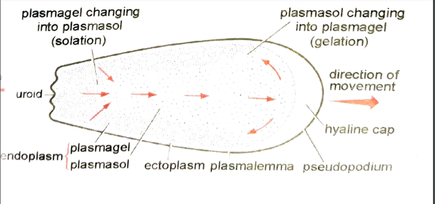
[e] Sol gel theory
- This theory was given by Hyman in 1917 and later supported by Pantin and Mast. It states that amoeboid movement is due change in consistency of cytoplasm. It is most widely excepted theory till date.
- It is also known as change in viscosity theory. It involves 4 process for amoeboid movement
- Plasamalemma is a outermost thin, elastic cell membrane become adhere to substratum.
- There is a local partial liquefication of the plasmagel at the interior end. That causes the central plasmasol under tension to flow forward against this weaken area to produce a bulge, the beginning of pseudopodium. It rapidly changes into plasmagel around periphery. Thus forming a gelatinized tube within which the plasmagel continue to flow forward.
- Inner plasmagel interiorly undergo solation which allow to maintain constant flow of plasmasol from behind in the direction of movement.
- Contraction of elastic plasmagel tube at which is located on outer side moves from in front backward while main bulk of body moves forward. The plasmagel thus exert squeezing motion from the side and near the amoeba forcing the plasmasol ahead. At tip of pseudopodium the endoplasm is changed to ectoplasm.
[f] Folding and unfolding theory
- This theory was proposed by Goldacre and Lorch folding and unfolding of protein chain leads to contraction and relaxation of protein molecule. They suggested that sol state of protoplasm is due to folding of protein whereas gel state is to unfolding of protein. Folding leads to contraction of end and amoeba progress on the other hand unfolding leads to liquefied sol state which is forced by central endoplasm and pushed forward. Considerable amount of ATP is invested in the form of energy for folding and unfolding of protein.
[g] Front and fountain zone contraction theory
- It was proposed by Allen in 1961. He compared amoeboid movement with muscle movement where contraction takes place. Where protein contraction leads to endoplasm contraction anterior end so that amoeba moves forward. The endoplasm is constantly converted to ectoplasm anteriorly and ectoplasm to endoplasm posteriorly.
[h] Reversible gel-sol transformation theory.
- Given by Yagi and Marsland this theory is most accepted theory explanation of amoeboid movement. This theory suggest that solation at anterior end occur into which endoplasm flows under pressure generated by contraction of the cortical plasmagel at the posterior end. This results in propulsion of amoeba
FLAGELLAR MOVEMENT
Single long locomotory flagella is enough for euglena movement. During swimming the flagella is directed obliquely backward toward the side bearing stigma. It undergo spiral undulations with waves that are transmitted from the base to the tip cuasing beating or sideway lashing. It beats on an average of 12 beats per second. This beating of flagellum drives the water backward and allow whole body to move forward. Each beat not only allow the body to move forward but also to one side. Hence when the body repeats one type of movement over and over the organism revolve in circle or gyrates.
As it directed to the backward direction to the long axis of the body the organism also rotate on its axis. It has been now calculated that euglena rotate at 1 turn per second speed.

CILLIARY MOVEMENT
Paramecium has streamlined body which enable it swim about in water with a minimum amount of friction.
[a] Cilliary beats
- During movement, a cilium oscillates like a pendulum. Each oscillation comprises fast and effective stroke and a slow recovery stroke. During effective stroke cilia becomes slight curve and rigid and strikes the water like a oar, so that body is propelled in opposite direction of stroke.
- At the cilia of body do not move simultaneously and independently but progressively in a characteristic wave like manner called metachronal rhythm. The cilia in longitudinal row beat in characteristic wave beginning at the anterior end. Cilia in longitudinal row beats one behind other where as in transverse row move synchronously.
[b] Mode of swimming
- They rotate spirally along the left handed helix.
- The body do not move directly backward but somehow how tilt to the right
- Secondly the cilia of oral groove strike obliquely and more vigorously so as to turn anterior end continuously away from the oral side and move in circle. The combine action causes the movement of animal along the fairly straight path rotating about its axis in an anti clockwise direction.
- In backward movement paramecium follow the straight path. This is due to the fact that the effective stroke is carried out anteriorly.
![FIGURE DEPICTING CILLIARY MOVEMENT [A] EFFECTIVE STROKE [B] RECOVERY STROKE](https://www.zoologytalks.com/wp-content/uploads/2021/11/1-9-300x183.png)
CONCLUSION
- There are large number of protozoans which moved with the help of or more whipped like structure called flagella. The movement can also takes place with help of cilia or with the help of pseudopodia in case of amoeba. Most unicellular organisms are included in this catergory. The flagellates are either plant like typically having chloroplast or animal like with no chloroplast.
- Protozoa that moves with the help of cilia are refer as ciliates and are included in subphylum cilliophora. Beside the character of these locomotor organ all ciliates posses two nuclei macro and micro nuclei.
- Amoeba is most popular free living protozoa it is regarded as a lowest form of animal as it body consist of mere spaces of protoplasm. It serves as an interesting and suitable material for laboratory as it is easy to obtain and very slow in locomotion. Amoeba perform locomotion with the help of pseudopodia various theory are given to describe its motion.
Share this:

Discover more from ZOOLOGYTALKS
Subscribe to get the latest posts sent to your email.