
DISSSECTION OF PILA GLOBOSA (APPLE SNAIL)
Pila Globosa is the most common and abundant gastropod, inhabiting ponds, rivers, tanks and rice-fields. It is very familiar for dissection and is also famous for its very slow movement called as snail pace.
External features of Pila Globosa (Apple Snail) with shell and operculum Procedure :

For studying external features, take formalin-preserved specimens. Note the following :-
- Shell: It is a univalve globose type. It is spirally coiled around a central axis called as columella.
- Apex : It forms the first part of the shell, situated at the top of the spiral. It is also called as protoconch .
- Whorls : The body coils of the shell are known as whorls and they are in open communication. The coils gradually increase from apex. Immediately after apex is penultimate whorl and then body whorl. The body whorl is the largest and successive whorls are demarcated by sutures.
- Varices : The surface of the shell is marked lines by numerous lines of growth, called as varices. of growth
- Columella: It is the central axis around which or varices shell is coiled.
- Mouth or aperture : The body whorl opens to outside by mouth or aperture.
- Peristome : The smooth and continuous margin of the aperture is called as peristome.
- Outer lip : It forms outer margin of the mouth. apex of shell
- Columellar lip : It forms inner lip.
- Umbilicus : The hollow tube-like columella opens to outside by umbilicus.
- Dextral shell : The shell of Pila shows clockwise spiralling, called as dextral shell.
- Operculum: The aperture of the shell is closed by operculum which is attached tightly to the hinder part of the foot.

Soft parts Procedure of Pila Globosa (Apple Snail)
For studying soft parts, break the body whorl nearly up to first suture. Keep operculum on the lower side. To study external features of soft parts remove the shell completely. The body of the Pila Globosa, After the shell is removed, is differentiated into distinct regions.
- Head: It is anterior fleshy part of the body overhanging foot. It is produced anteriorly into the contractile snout, which bears mouth and 2 pairs of tentacles. First pair is smaller, while second pair of tentacles is larger. At the base of each second pair tentacles is a stalked eye or ommatophore.
- Foot: It is large, strongly muscular, ventral part of the body. In fully expanded condition, foot shows triangular appearance. The part of the foot bearing operculum dorsally is called as operculiferous lobe. When foot is withdrawn, the operculum completely fits into the mouth.
- Visceral mass: It constitutes a sort of hump on the dorsal side called as visceral hump. It is spirally coiled according to the coiling of the shell.
- Mantle: The skin of the visceral mass forms a thin and delicate covering called as mantle or pallium. Mantle encloses a large cavity in dorso-Iateral position called as mantle cavity or pallial cavity.
General anatomy of Pila Globosa (Apple Snail) Procedure :
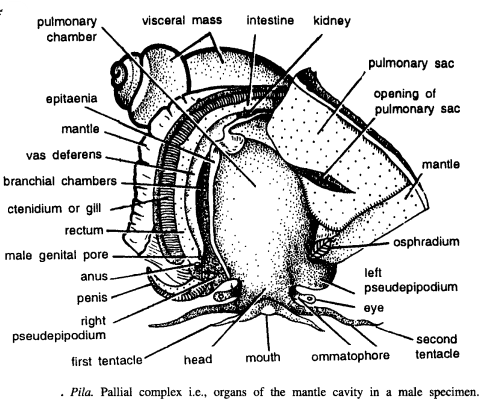
For general anatomy, do not remove animal from the shell. Break the shell up to first suture. Keep the snail with visceral hump upwards. Make an oblique incision from the left side along the pigmented area of the mantle. Reflect the mantle flap and pin it up. Study the following parts
- A. Mantle cavity and pallial complex :- Mantle encloses a large dorso lateral cavity, called as mantle cavity or pallial cavity. Animal retracts its head into this cavity. The pallial cavity encloses the following organs, collectively called as pallial complex.
- Right and left nuchal lobes : At the sides of the head and over the foot, the mantle is prolonged into highly contractile and fleshy nuchal lobes or pseudepipodia
- Respiratory siphon : The left nuchal lobe forms respiratory siphon.
- Epitaenia: From the anterior edge of right nuchal lobe a prominent ridge or raised area extends up to extreme posterior end and is called as epitaenia. It divides mantle cavity into right branchial chamber and left pulmonary chamber.
- Organs of the branchial chamber
- Ctenidium: Monopectinate. Hangs freely vertically downwards from the dorso-Iateral wall of the mantle cavity.
- Rectum: Rectum lies in front of the ctenidium.
- Genital duct: Male or female genital duct lies close to the rectum. In male, penis arises from the mantle edge.
- Organs of pulmonary chamber
- Pulmonary sac : It is large bag-like structure, hanging down from the roof of the mantle cavity.
- Osphradium: It arises from the mantle and is found adjacent to left nuchal lobe.
Nervous system of Pila Globosa (Apple Snail) Procedure

Take a nicely preserved animal. Break the body whorl up to suture. Cut the mantle fold obliquely and remove the fold by cutting it posteriorly carefully so as not to destroy the visceral ganglia. The pulmonary chamber is exposed and buccal mass can be easily seen. Remove the muscular covering over buccal mass very carefully and under the muscular coat thick cerebral commissure will be seen. On sides, cerebral ganglia are found Trace supra-intestinal ganglion posterior to left nuchal lobe.
Visceral ganglia are found in posterior region. The supra-intestinal nerve is a very thin nerve passing over gut and going deep to join the right pleuropedal ganglionic mass. Cut the gut part and remove buccal mass carefully to expose complete nervous system. Expose the following ganglia, connectives and commissures
- Ganglia
- Cerebral ganglia : A pair of these ganglia, situated on dorso-Iateral sides of the buccal mass and triangular in shape.
- Buccal ganglia: A pair of small triangular ganglia dorso-Iaterally situated at the junction of buccal mass and oesophagus.
- Pleuro-pedal ganglia : A pair of ganglionic mass, lying on either ventro-Iateral side of the buccal mass. Pleural outer one and pedal inner one are fused together to form pleuro-pedal ganglionic mass.
- Supra-intestinal ganglion: Unpaired fusiform ganglion lying in a sinus behind the left pleuro-pedal ganglionic mass.
- Visceral ganglia: Found in the lower end of the visceral mass.
- Connectives
- Two cerebro-pleural connectives : They connect cerebral and pleural ganglia on either side.
- Two cerebro-pedal connectives : They connect cerebral and pedal ganglia on either side.
- Two cerebro-buccal connectives : They connect cerebral and buccal ganglia.
- Infra-intestinal visceral connective : It connects right pleuro-pedal ganglia to visceral ganglia.
- Supra-intestinal pleural connective : It connects supra-intestinal ganglion to right pleuro-pedal ganglia.
- Infra-intestinal connective: It connects left pleuro-pedal ganglia to infra-intestinal ganglion fused with right pleuro-pedal ganglionic mass.
- Commissures
- Cerebral commissure connects the two cerebral ganglia.
- Pedal commissure connects the two pedal ganglia.
- Buccal commissure connects the buccal ganglia.
Share this:

Discover more from ZOOLOGYTALKS
Subscribe to get the latest posts sent to your email.