Introduction
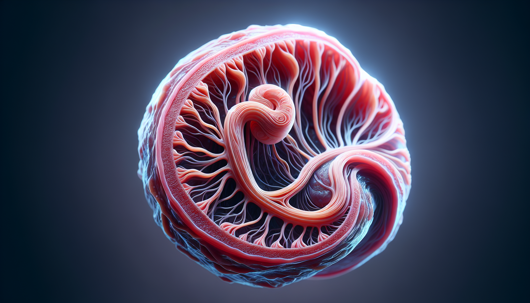
Epitheliochorial Placenta Structure and Function- The epitheliochorial placenta is a unique and significant type of placental structure found in certain mammals, particularly ungulates like horses, pigs, and some primates. This form of placenta is crucial in understanding reproductive biology, maternal-fetal interactions, and the evolutionary adaptations that different species have developed for successful reproduction. In this article, we delve into the intricacies of the epitheliochorial placenta, exploring its structure, function, and relevance in the broader context of mammalian reproduction.
What is the Epitheliochorial Placenta?

The placenta is an essential organ that forms during pregnancy, facilitating the exchange of nutrients, gases, and waste between the mother and the developing fetus. In mammals, the placenta can vary significantly in its structure and the degree of intimacy between maternal and fetal tissues. The epitheliochorial placenta is characterized by a relatively simple structure, where the maternal and fetal blood supplies are separated by multiple tissue layers.
In the epitheliochorial placenta, there are six distinct layers of tissue separating the maternal blood from the fetal blood. These layers include:
- Maternal Endothelium: The innermost lining of the maternal blood vessels.
- Maternal Connective Tissue: The supportive tissue surrounding the maternal blood vessels.
- Maternal Epithelium (Endometrium): The outermost layer of the maternal tissue, also known as the endometrial epithelium.
- Fetal Trophoblast: The outermost layer of fetal cells that are in contact with the maternal tissues.
- Fetal Connective Tissue: The supportive tissue surrounding the fetal blood vessels.
- Fetal Endothelium: The innermost lining of the fetal blood vessels.
These layers ensure that the maternal and fetal blood supplies do not directly mix, which is a defining feature of the epitheliochorial placenta. The separation by these layers makes the epitheliochorial placenta less efficient in nutrient and gas exchange compared to other placental types, such as the haemochorial placenta found in humans and rodents.
Species with Epitheliochorial Placentas
The epitheliochorial placenta is commonly found in certain groups of mammals, particularly:
- Ungulates: This includes horses, pigs, and some ruminants like cattle and sheep.
- Lemurs: Among primates, lemurs are known to have an epitheliochorial placenta.
- Whales and Dolphins: Cetaceans also exhibit this type of placentation.
These species have evolved the epitheliochorial placenta as a reproductive adaptation that suits their unique ecological niches and life histories.
Structural and Functional Characteristics
One of the key features of the epitheliochorial placenta is the presence of the maternal epithelium, which remains intact and forms a barrier between the mother and the fetus. This structural arrangement has several implications for the function of the placenta:
- Reduced Immune Interaction: The multiple layers of separation reduce the likelihood of maternal immune cells recognizing and attacking the fetal tissues as foreign. This is particularly important in species with long gestation periods, where maintaining immune tolerance is critical.
- Controlled Nutrient Transfer: The presence of six tissue layers means that nutrient and gas exchange between the mother and fetus is more controlled and less efficient compared to other placental types. This necessitates longer gestation periods and may influence the birth weight and development of the offspring.
- Protection Against Pathogens: The layers of tissue provide a protective barrier against potential pathogens, reducing the risk of infections passing from the mother to the fetus. This is an important advantage for species that may be exposed to various environmental pathogens.
- Evolutionary Adaptations: The epitheliochorial placenta is believed to be an evolutionary adaptation to specific environmental conditions and reproductive strategies. For example, in ungulates, which often give birth to relatively large, well-developed offspring, the epitheliochorial placenta supports prolonged gestation and slow, steady nutrient transfer.
Comparative Analysis with Other Placental Types
To better understand the epitheliochorial placenta, it is useful to compare it with other types of placentation found in mammals:
- Haemochorial Placenta: Found in humans, rodents, and some other mammals, the haemochorial placenta has fewer tissue layers separating the maternal and fetal blood supplies. This allows for more efficient nutrient and gas exchange but also increases the risk of immune interaction and pathogen transmission.
- Endotheliochorial Placenta: Found in carnivores like dogs and cats, the endotheliochorial placenta has four layers, with the maternal endothelium in direct contact with the fetal trophoblast. This type of placenta offers a balance between nutrient transfer efficiency and protection.
- Syndesmochorial Placenta: Found in ruminants like cows and sheep, the syndesmochorial placenta is similar to the epitheliochorial placenta but with some maternal tissue layers eroded, allowing for closer contact between maternal and fetal tissues.
The differences in these placental structures highlight the diversity of reproductive strategies among mammals and the role of the placenta in facilitating successful reproduction under varying ecological conditions.
Significance in Reproductive Biology
The study of the epitheliochorial placenta provides valuable insights into reproductive biology, particularly in understanding how different species have evolved to maximize reproductive success. The epitheliochorial placenta’s structure reflects a trade-off between protecting the fetus from maternal immune responses and pathogens while ensuring sufficient nutrient and gas exchange during gestation.
For veterinarians, animal breeders, and conservationists, knowledge of the epitheliochorial placenta is crucial for managing the reproductive health of species that exhibit this type of placentation. It informs breeding programs, helps in diagnosing and treating reproductive disorders, and contributes to the conservation of endangered species by improving our understanding of their reproductive physiology.
Conclusion
The epitheliochorial placenta is a fascinating example of the diversity of placental structures in mammals. Its unique characteristics, including the multiple layers of tissue separating maternal and fetal blood, make it an essential area of study in reproductive biology. By understanding the epitheliochorial placenta, researchers and practitioners can gain deeper insights into the reproductive strategies of different species and the evolutionary adaptations that have shaped their reproductive success.
Discover more from ZOOLOGYTALKS
Subscribe to get the latest posts sent to your email.


